Reading protein–lipid binding in real time
Almost every protein–lipid assay answers “how tightly?” Very few answer “how fast, and how long?” In a cell those are not the same question. Getting at the second one took a long time on buffer conditions.
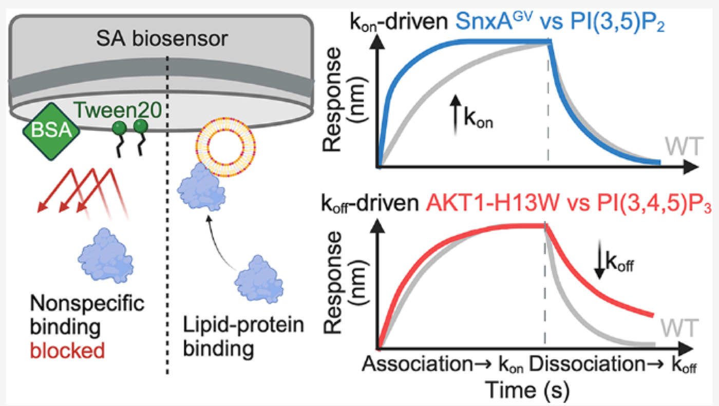
Affinity does not describe the whole binding process
A eukaryotic cell keeps its compartments straight partly by chemistry. Phosphoinositides, or PIPs, the eight phosphorylated derivatives of phosphatidylinositol, are distributed unevenly across the endomembrane system: PI(4,5)P₂ marks the plasma membrane, PI(3)P early endosomes, PI(3,5)P₂ late endosomes. Proteins with lipid-binding domains (PH, PX, ENTH, GRAM, C2 …) read that distribution and go where they are needed. A single misread can be pathological: the E17K mutation in the AKT1 PH domain changes which PIP the domain prefers and drives cancer.
When we want to describe one of these interactions, we almost always reach for a single number: the dissociation constant, KD. It is a useful number, and it is also a ratio:
KD = koff / kon
Two proteins can share a KD of 10 nM and behave quite differently in a cell. One reaches the membrane fast and leaves fast; the other takes longer to find the target and then stays for minutes. If the protein's job is to respond to a brief PIP pulse, those two behaviours have different consequences. KD on its own does not show the difference.
Why liposomes make this hard
Bio-layer interferometry (BLI) is, in principle, the right instrument for this. A biosensor tip is dipped into a well; molecules binding at the tip surface change the interference pattern of reflected white light, and you watch the response in nanometres, live. Dip into protein and you get association; dip into buffer and you get dissociation. Fit both, and you have kon and koff directly.
The protein adsorbs onto the sensor on its own: onto the streptavidin layer, the sensor matrix, exposed biotin. That happens whatever you are measuring, and the instrument records it alongside the binding you want. Background can be half your signal, and it carries kinetics of its own, which contaminate the fit.
The usual fix is detergent. With a protein, a small molecule or a nucleic acid target it costs almost nothing. With liposomes it does cost something: the concentration that strips adsorbed protein is usually enough to dissolve the liposome as well.
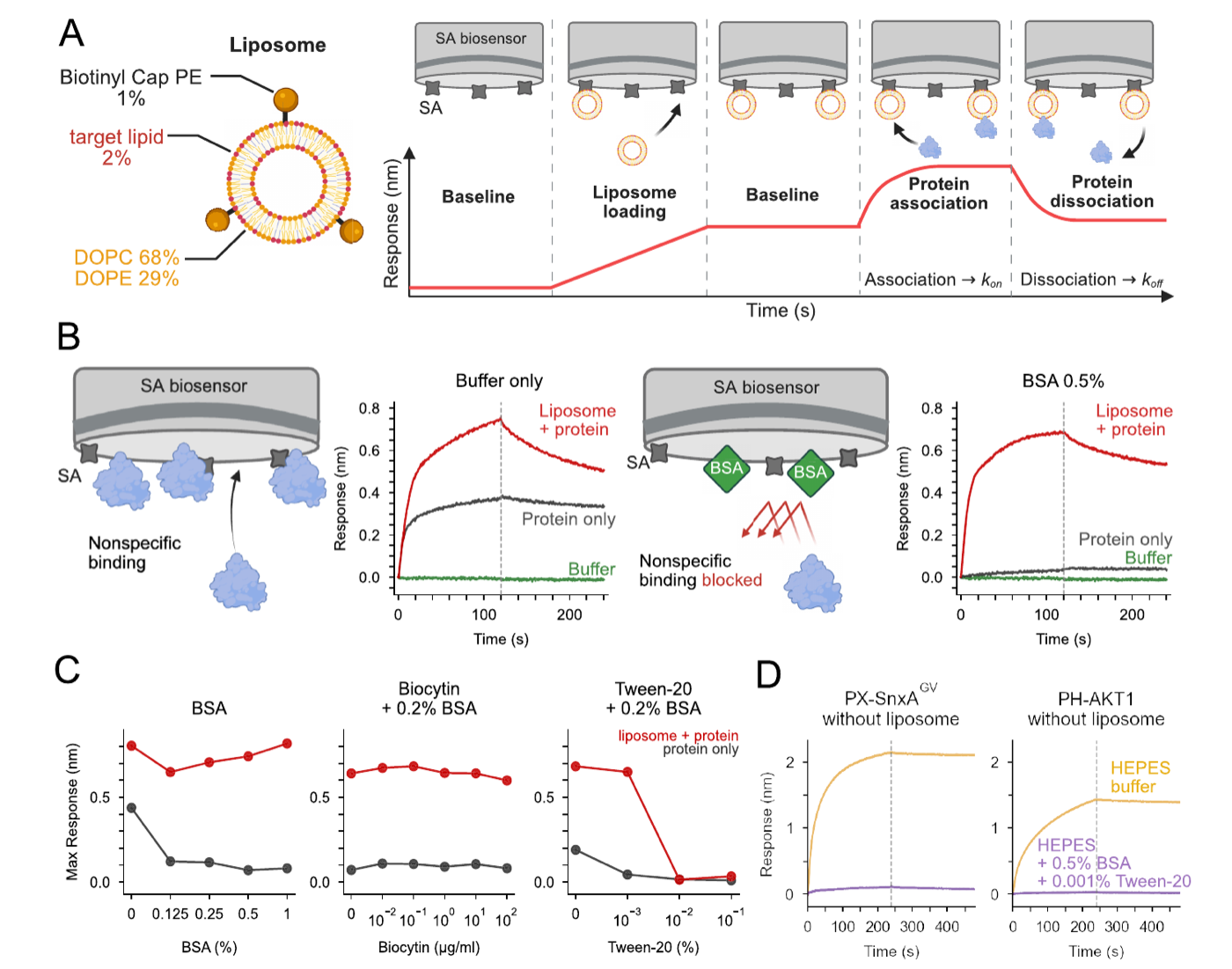
Which additives worked
We screened three additives against both signals at once, the specific one we wanted to keep and the nonspecific one we wanted to remove. The working conditions sit inside a narrow window:
- BSA at 0.5% does most of the work. It coats the sensor surface and blocks protein–surface contact. Specific binding is untouched.
- Biocytin, the obvious candidate for capping free streptavidin sites, did essentially nothing. We reported that negative result because it is the reagent people reach for first.
- Tween-20 at 0.001% removes the last of the background. At 0.01% and above, the specific signal collapses too, because the liposome itself comes apart.
Does it recover known biology?
We ran a specificity panel first: three well-characterised domains against a set of liposomes covering PI, PI(3)P, PI(3,4)P₂, PI(3,5)P₂, PI(4,5)P₂ and PI(3,4,5)P₃, plus binding-altered mutants of each.
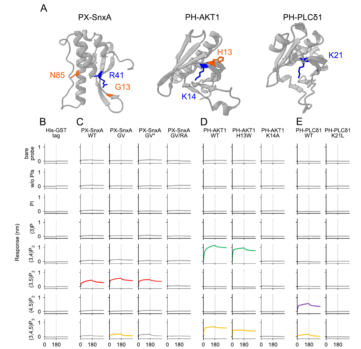
Everything landed where the literature says it should, and the affinity-abolishing mutants gave no signal. With that in hand we were willing to use the assay on targets nobody had measured.
What the rate constants revealed
We took concentration series across each pair and fitted them globally. Two of the mutant series moved in opposite directions.
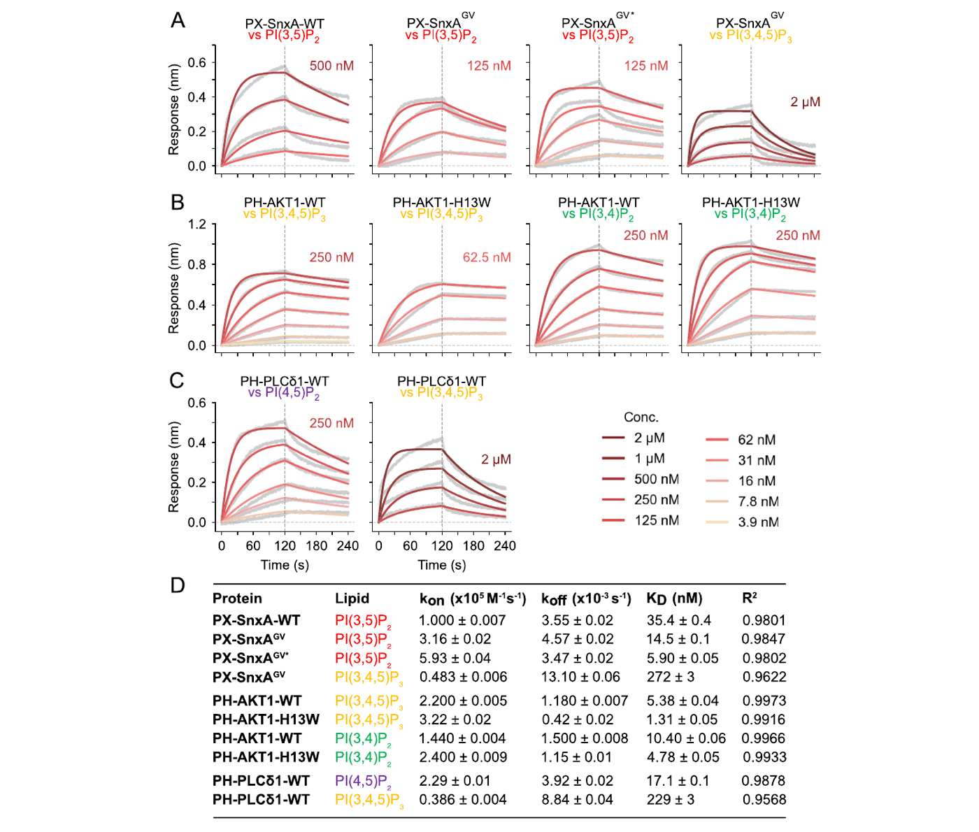
In the PX-SnxA series, affinity improves about six-fold from wild type to the double mutant, and kon rises six-fold while koff barely moves. These mutations make the domain find and engage the membrane faster. They do not make the bound complex more stable.
In the AKT1 series, H13W improves affinity roughly four-fold, and it gets there mostly by cutting koff by a factor of three. This mutation stabilises the complex once formed.
In an equilibrium assay both read as “tighter binding”. The mechanisms differ: one captures faster, the other stays bound longer. For a domain that has to respond to a brief lipid signal, those two have different consequences in a cell.
If you are going to measure protein–lipid kinetics by BLI
- The main source of nonspecific binding is probably protein adsorbing onto the probe surface.
- BSA is probably the most effective blocker. The paper used 0.5%; push it higher if you need to, to 1% for instance.
- Biocytin may do very little. Streptavidin is probably not the main source of nonspecific binding.
- Tween-20 may help, but raise the BSA concentration first: the safe window for Tween-20 is narrow.
- A liposome surface carries a great many binding sites, so treat the fitted parameters with care. Raising the target-lipid fraction does give a larger binding signal, but take that at face value cautiously: avidity, or poor diffusion, can produce the same appearance.
- A GST tag helps solubility, but GST dimerises, so a result obtained that way cannot be reported as clean 1:1 kinetics.
Source paper. Yao, P.; Nishimura, T.*; Tsuboyama, K.* Quantitative Kinetic Analysis of Protein–Phosphoinositide Binding by Optimized Liposome-Based Bio-Layer Interferometry. Biochemistry 2026, special issue “Lipids and Lipidation”. doi.org/10.1021/acs.biochem.6c00344
Figures are reproduced from that article, of which I am the first author. Please cite the paper rather than this page.
实时读出蛋白质与脂质的结合
几乎所有蛋白质–脂质实验回答的都是「结合有多紧」,很少有人回答「结合得多快、停留多久」。在细胞里这不是同一个问题。为了测第二个,我们在缓冲液条件上花了很长时间。
亲和力不能完整描述结合过程
真核细胞靠化学把自己的各个区室区分开来。磷脂酰肌醇(PIPs,即磷脂酰肌醇的 8 种磷酸化衍生物)在内膜系统中分布并不均匀:PI(4,5)P₂ 标记质膜,PI(3)P 标记早期内体,PI(3,5)P₂ 标记晚期内体。带有脂质结合结构域(PH、PX、ENTH、GRAM、C2 ……)的蛋白质读取这套分布,从而知道该去哪里。读错一次就可能致病:AKT1 PH 结构域的 E17K 突变改变了它偏好的 PIP 种类,进而驱动癌症。
要描述这样一个相互作用,我们几乎总是用同一个数字:解离常数 KD。它有用,同时也是一个比值:
KD = koff / kon
两个蛋白的 KD 可以同样是 10 nM,在细胞里的行为却差很多。一个很快到达膜,也很快离开;另一个要花时间才找到目标,但会停留几分钟。如果这个蛋白要响应的是一次短暂的 PIP 脉冲,这两种行为的后果不一样。只报 KD 看不到这层差别。
脂质体让这件事变难
生物层干涉法(BLI)原则上正是合适的工具。传感器针尖浸入孔中,结合到针尖表面的分子改变反射白光的干涉图样,你就能以纳米为单位实时看到响应。浸入蛋白溶液即为结合,浸入缓冲液即为解离。两段都拟合,就直接得到 kon 与 koff。
蛋白自己就会吸附到传感器的各个部位:链霉亲和素层、传感器基质、暴露出来的生物素。无论你测的是什么,这层吸附都在,仪器会把它和你想要的信号一起记下来。背景可以占到信号的一半,它自己也有动力学,会污染拟合。
常规解法是加去污剂。测蛋白、小分子、核酸的时候,这一步几乎没有代价。换成脂质体就有代价了:能洗掉吸附的去污剂浓度,往往也足以把脂质体溶掉。
哪些添加剂有效
我们把三种添加剂同时对两个信号做了筛选:想保留的特异信号,和想去掉的非特异信号。有效的条件落在一个很窄的窗口里:
- 0.5% BSA 起主要作用。它覆盖传感器表面,挡住蛋白与表面的接触,特异结合不受影响。
- Biocytin 用来封闭游离的链霉亲和素位点,基本没有作用。它常常是大家第一个想到的试剂,所以这个阴性结果我们也写进了论文。
- 0.001% Tween-20 清掉最后一点背景。到 0.01% 及以上,特异信号也一起塌了,因为脂质体本身被破坏了。
它能重现已知的生物学吗
我们先做了特异性验证:三个被充分研究过的结构域,对一组覆盖 PI、PI(3)P、PI(3,4)P₂、PI(3,5)P₂、PI(4,5)P₂ 与 PI(3,4,5)P₃ 的脂质体,外加各自的结合改变突变体。
结果都落在文献报道的位置上,破坏亲和力的突变体也没有信号。这一步做完,我们才有把握把它用在没人测过的对象上。
动力学参数揭示了新的发现
我们对每一对做了浓度梯度,然后做全局拟合。有两个突变体系列的走向恰好相反。
看 PX-SnxA 这一组:从野生型到双突变体,亲和力提高约 6 倍,而kon 提高了 6 倍,koff 几乎没动。这些突变让结构域更快地找到并接触膜,却并没有让已形成的复合物更稳定。
再看 AKT1 系列:H13W 让亲和力提高约 4 倍,而它主要是靠把 koff 降到三分之一做到的。这个突变稳定的是形成之后的复合物。
在平衡实验里,这两种情况都只表现为「结合更紧了」。但机制不同:一个是捕获更快,一个是停留更久。如果这个结构域要响应的是一次瞬时的脂质信号,这两种机制在细胞里的后果并不一样。
如果你也要用 BLI 测量蛋白和磷脂的结合动力学
- 非特异性结合的主要来源,可能是蛋白与探针表面的吸附。
- BSA 应该是最有效的封闭剂。论文里用的是 0.5%,必要时可以往上加,比如 1%。
- Biocytin 的封闭效果可能很有限。链霉亲和素未必是非特异结合的主要来源。
- Tween-20 可能有效,但优先考虑提高 BSA 浓度:Tween-20 的安全浓度窗口很窄。
- 脂质体表面遍布结合位点,拟合出来的参数要仔细斟酌。提高目标脂质的比例确实会带来更高的结合信号,但不要盲目乐观,那可能是 avidity 效应或者扩散效率不足造成的假象。
- GST 标签有助溶效果,但 GST 自身会形成二聚体,因此这样得到的结果不能当作干净的 1:1 结合动力学来报告。
原始论文。Yao, P.; Nishimura, T.*; Tsuboyama, K.* Quantitative Kinetic Analysis of Protein–Phosphoinositide Binding by Optimized Liposome-Based Bio-Layer Interferometry. Biochemistry 2026,专刊 “Lipids and Lipidation”。doi.org/10.1021/acs.biochem.6c00344
文中图片转载自该论文,本人为第一作者。引用请引原文,而非本页。
タンパク質と脂質の結合をリアルタイムで読む
タンパク質と脂質の結合を測るアッセイの多くは、結合の強さしか答えてくれない。どれだけ速く結合し、どれだけ長く留まるのかを答えるものは少ない。細胞の中では、この二つは別の問いである。後者を測れるようにするまでに、緩衝液の条件をずいぶん長く探すことになった。
親和性だけでは結合の過程は分からない
真核細胞は、区画ごとの化学組成によって自分の内部を区別している。ホスホイノシチド(PIPs)はホスファチジルイノシトールがリン酸化された 8 種類の脂質で、細胞内膜系に一様には分布しない。PI(4,5)P₂ は細胞膜、PI(3)P は初期エンドソーム、PI(3,5)P₂ は後期エンドソームの目印になる。脂質結合ドメイン(PH、PX、ENTH、GRAM、C2 など)を持つタンパク質はこの分布を読み取り、自分が働くべき場所へ向かう。読み違えが病気につながることもあり、AKT1 の PH ドメインに入る E17K 変異は、このドメインが好む PIP の種類を変えてがんを引き起こす。
この種の相互作用を記述するとき、持ち出される数値はたいてい一つ、解離定数 KD である。有用な数値だが、これは比でもある。
KD = koff / kon
KD が同じ 10 nM でも、細胞の中での振る舞いは大きく変わりうる。速く膜に着いて速く離れるものもあれば、標的を見つけるまでには時間がかかるが、いったん結合すれば数分間留まるものもある。短い PIP シグナルに応答するのがそのタンパク質の役割なら、この二つが細胞にもたらす結果は同じではない。KD だけを見ていても、その差は見えてこない。
リポソームだと話が難しくなる
バイオレイヤー干渉法(BLI)は、原理から見ればこの測定に向いた装置である。バイオセンサーの先端をウェルに浸すと、先端の表面に結合した分子が反射光の干渉パターンを変える。その変化をナノメートル単位でそのまま追えるので、タンパク質溶液に浸せば結合が、緩衝液に浸せば解離が見える。両方をフィットすれば kon と koff がそのまま求まる。
やっかいなのはタンパク質自身である。タンパク質はセンサーのあちこちに吸着する。ストレプトアビジン層、センサーマトリックス、露出したビオチン。何を測っていても起こることで、装置は目的のシグナルと区別せずに記録してしまう。バックグラウンドがシグナルの半分に達することもあり、しかもそれ自体が速度論的に振る舞うので、フィットが歪む。
ここで普通は界面活性剤を入れる。相手がタンパク質や低分子、核酸であれば、この一手にほとんど代償はない。リポソームではそうもいかない。吸着を落とせる濃度は、たいていリポソームそのものを溶かしてしまう濃度でもある。
効いた添加剤
三つの添加剤について、残したい特異的なシグナルと消したい非特異的なシグナルの両方を同時に見ながら条件を振った。使える条件の幅は狭かった。
- 0.5% BSA が中心的に効く。センサー表面を覆ってタンパク質が表面に触れるのを防ぐ一方、特異的な結合のほうは妨げない。
- Biocytin は遊離のストレプトアビジン部位を塞ぐつもりで使ったが、ほとんど効果がなかった。最初に思いつく試薬なので、この陰性の結果も論文に載せてある。
- 0.001% Tween-20 で残りのバックグラウンドが消える。0.01% 以上にすると特異的なシグナルまで落ちるが、これはリポソーム自体が壊れているためである。
既知の生物学を再現できるか
まず特異性を確かめた。よく調べられている三つのドメインを、PI、PI(3)P、PI(3,4)P₂、PI(3,5)P₂、PI(4,5)P₂、PI(3,4,5)P₃ を含むリポソームのパネルにかけ、あわせて結合の変わる変異体も測った。
どれも文献どおりの位置に落ち、親和性を失う変異体はシグナルを出さなかった。ここまで確かめたので、まだ誰も測っていない対象にこのアッセイを使うことにした。
速度定数が明らかにしたこと
それぞれの組み合わせについて濃度系列を取り、グローバルにフィットした。変異体の二つの系列が、互いに逆の方向へ動いていた。
PX-SnxA の系列では、野生型から二重変異体にかけて親和性が約 6 倍上がる。このとき kon が 6 倍になる一方、koff はほとんど変わらない。変異によって膜を見つけて接触するまでが速くなっているのであって、できあがった複合体が安定になっているわけではない。
AKT1 の系列では、H13W が親和性を約 4 倍上げる。こちらは koff が 1/3 に下がることによるもので、できた複合体のほうが安定化している。
平衡アッセイでは、どちらも「結合が強くなった」としか見えない。しかし機構は違う。一方は捕捉が速くなり、もう一方は滞在が長くなる。短い脂質シグナルへの応答が役割のドメインであれば、この二つが細胞にもたらす結果は同じではない。
BLI でタンパク質と脂質の結合速度論を測るなら
- 非特異的結合の主な出どころは、おそらくタンパク質がプローブ表面に吸着することである。
- BSA が最も効くブロッカーだと思う。論文では 0.5% を使ったが、必要なら 1% 程度まで上げてよい。
- Biocytin によるブロッキングはあまり効かないかもしれない。ストレプトアビジンが非特異的結合の主な出どころとは限らない。
- Tween-20 も効く可能性はあるが、先に BSA の濃度を上げるほうを試したい。Tween-20 は使える濃度の幅が狭い。
- リポソームの表面には結合部位がいくらでもあるので、フィットで得たパラメータは慎重に扱ったほうがよい。標的脂質の割合を上げれば結合シグナルは大きくなるが、そのまま強く結合していると解釈しないほうがよい。avidity 効果や拡散の遅さが、同じ見え方をつくることがある。
- GST タグは可溶化には役立つ。ただし GST 自体が二量体をつくるので、こうして得た結果を 1:1 の結合速度論として報告することはできない。
原論文。Yao, P.; Nishimura, T.*; Tsuboyama, K.* Quantitative Kinetic Analysis of Protein–Phosphoinositide Binding by Optimized Liposome-Based Bio-Layer Interferometry. Biochemistry 2026、特集号 “Lipids and Lipidation”。doi.org/10.1021/acs.biochem.6c00344
図は同論文からの転載で、筆頭著者は私です。引用は本ページではなく原論文へお願いします。